CPT Coding Tips - 96125 (Standardized Cognitive Performance Testing, Per Hour)

Log this code when:

  1. The combined time it takes to conduct the evaluation, interpret the results, and write the evaluation report* is at least 31 minutes to report the first hour, 91 minutes to report the second hour, and so on.
  2. The test is completed using a standardized assessment, independently or in conjunction with subjective observations and findings.

*Note: Clinicians may count interpretation and documentation time toward the minimum minutes only when billing for 96125, and only for Medicare Part B patients. Medicare Part A minutes still follow RAI manual guidelines of direct face-to-face time, which is followed regardless of code definition. Additionally, when administered as the initial evaluation, this code is non-MDS for Part A payers.

Completing standardized assessments supports evidence-based practice and helps to clearly identify where to target intervention for the best results. While tools like the SLUMs offer insight as to where a deficit may be occurring, they only allow a general categorization of cognitive impairment: normal, mild, or severe/dementia.

Utilizing formal standardized assessments for cognition will help determine which component of the cognitive impairments need intervention. With so many components of cognition, it’s best to assess as many areas as possible. Cognition is the greatest predictor of function. The more areas assessed, the stronger the plan of care and better patient outcomes.

Please refer to the Cognitive Performance Assessment POSTette for additional information.

Importance of Therapy Intervention for Patients Post-COVID

By Tamala Sammons, MA, CCC/SLP, Therapy Resource
COVID-19 is a respiratory viral disease with multi-organ involvement resulting in potentially temporary and episodic health challenges such as impaired lung function, physical deconditioning, cognitive impairments, impaired swallow and communication, and mental health disorders.

The effect of the virus on the respiratory system appears to range from a simple respiratory tract infection to acute respiratory distress syndrome (ARDS) with multi-organ failure.
People with COVID-19 may also develop coagulopathy that can lead to venous thromboembolism (VTE) and microvascular thrombosis throughout the body, increasing the risk of negative mental health outcomes (https://academic.oup.com/ptj/article/100/12/2127/5903663).

Older adults respond to their own stress and that experienced by staff, which can increase the risk of behavioral problems. Isolation and lack of stimulation may also lead to loneliness and depression. Each of these negative psychological outcomes has a significant impact on an individual’s immune system and the ability to fight infection.

An increasing number of patients recovering from COVID-19 are having lingering cognitive symptoms, including confusion and impaired executive functions, short-term memory issues, and learning difficulties. For those who had compromised cognitive-communication status before COVID-19, we need to be ready for ongoing exacerbation of symptoms.

New survey standards: Surveyors are advised to investigate any concerns as part of the focused infection control survey related to residents who have had a significant decline in condition. Emphasis is on decline with the resident’s condition, both physical and/or psychosocial.
Therapy Interventions must address the clinical changes in our patients from this pandemic.

Occupational Therapy Intervention/Strategies
● Measure the effects of cardiac function and respiration, including any increased oxygen and/or oxygen weaning with ADLs (respiratory rehab ADLs; OT COPD treatment protocol)
● Assess functional mobility — high fall risk
● Provide energy conservation and work simplification interventions
● Consider cognition, cognitive rehabilitation and occupational performance; and address cognitive impairments
● Address psychosocial (OT psychosocial interventions), mental health (AOTA OT’s role with mental health recovery), stress (HRV training), and coping-related PTSD or anxiety disorder post-COVID-19
● Community reintegration

Physical Therapy Intervention/Strategies
● Measure the effects of cardiac function and respiration, including any increased oxygen and/or oxygen weaning with physical performance (respiratory rehab physical exercise; PT COPD treatment protocol)
● Provide strength and mobility interventions — exercise prescription, fall risk, take longer to regain strength
o Note: For exercise prescription, go back to pulmonary rehab principles and exercise prescription for older adults and modify based on symptoms, vital signs and RPE
o Aerobic exercise, strength training, flexibility intervention
o It is critical that physical therapists are aware of the clinical implications of coagulopathy and the prevalence of venous thromboembolism (VTE) in patients diagnosed with and recovering from COVID-19 through the promotion of early mobility and physical activity
● Provide pain assessments and intervention
● Community reintegration as indicated
● Need for outpatient services

Speech-Language Pathology Intervention/Strategies
● Measure the effects of cardiac function and respiration, including any increased oxygen and/or oxygen weaning with communication and swallow (respiratory rehab breathing interventions and using resistive devices; SLP COPD treatment protocol)
● Assess effective communication as it relates to respiratory function
● Assess cognition (great opportunity to use comprehensive cognitive assessments), provide cognitive rehabilitation to address cognitive impairments
o Note: Studies are emerging that suggest some type of association between neurological symptoms and COVID-19. Symptoms like headache, stiff neck, and loss of taste and smell raise suspicion of central nervous system involvement.
● Assess swallow; airway protection and cough strength. Integrate resistive breathing devices for intervention.
● Assess for continued loss of taste and smell and how that impacts hydration and nutrition.
● Need for outpatient services.

Exercising Your Passion for Learning

By Kai Williams, Therapy Resource, Keystone East, TX
There are many times that we run across clinicians that have such a passion for integrating new and innovative clinical ideas. Some move with hesitancy, unsure if their idea will really gain traction. We say to those that are hesitant, please Exercise your Passion for Learning! Malathy Venkatesh, PhD, CCC-SLP, originally joined our Mason Creek facility in Katy, Texas, in 2019. She recently relocated to our Tempe post-acute facility located in Arizona in late 2020. From the very first time I met Malathy, our conversations revolved around the underutilization of speech therapy services with a goal on how to increase awareness around clinical programming. Malathy has spent the last six months introducing LTC patients to the Montessori Method. Below you will find details about the Montessori approach to dementia from an SLP perspective, along with some ideas on how SLPs can use this approach in the LTC setting with examples of materials and goals.

Applying the Montessori Principles in the Management of Dementia: Speech Language
Pathologist’s Perspective
Malathy Venkatesh PhD CCC-SLP, Tempe Post-Acute, Tempe AZ
Speech Language Pathologists (SLPs) are constantly seeking approaches, materials and activities that place less demand on an impaired and progressively declining cognitive system such as the semantic and episodic memory, and more on engaging the relatively preserved procedural memory, to improve safety and functions in individuals with dementia. One approach that maximizes the preserved skills and abilities such as recognition of stimuli and reading is the Montessori approach for ageing and dementia. This approach, derived from the pedagogical approach to child education, has been developed and implemented successfully in adult day care centers and long-term care (LTC) settings.

One of the important principles of the Montessori approach is to encourage independence in individuals with dementia by engaging them in meaningful and purposeful activities in a prepared environment. The Montessori approach recommends that SLPs evaluate and manage individuals with dementia in their natural environment such as their rooms, dining room and activity room in addition to the clinical setting. The communication performance in a quiet therapy room may differ greatly from their communication abilities in a dining or activity room where they must adapt to the background noise and disproportionate visual stimulation. Two of the most common environmental barriers that affect the communication and interaction ability of individuals with dementia is light and contrast. The recommended environmental modifications for these barriers include incorporating bright but non-glare lighting in therapy spaces and reading spaces, minimizing unevenly lighted spaces, and preparing the environment where the figure contrasts from the background in such a way that it stands out, draws attention and is easy to locate.

The Montessori approach also relies heavily on “cueing,” using explicit visual cues in addition to verbal cues to facilitate initiation, maintenance and completion of an activity, and directions, signs around the facility, and memory books. Both texts and pictures are utilized while developing visual cues. Lighting and contrasts are important factors to be considered while displaying the visual cues. They should be displayed in a way that facilitates ease of recognition and reading and must fulfill the purpose that it intends to achieve, for example, locating rooms in the building.

The Montessori approach taps on the relatively preserved ability of reading aloud by encouraging this skill as a pleasure activity utilizing dementia-friendly books. These books use large-print text with high-contrast pictures and illustrations. They are colorful and, more importantly, they are books designed for senior adults and adults with dementia. Reading books helps maintain language and reading skills, preserve pincer grasp when turning the pages, and maintain attention and focus. As shown in Figure 1, the environment is prepared ahead of the reading task by choosing a well-lit, quiet, distraction-free spot in the room. The book is mounted on a stand for easy reading and for page turning. Also notice that there are no shadows or glare on the book.

Figure 1: Dementia-friendly book (Courtesy of Susan Ostrowski, Reading2Connect at
www.Reading2Connect.com)

Another example applying the Montessori philosophy for a simple verbal expression task is utilizing a picture cube and wh-questions cue cards as illustrated in Figure 2. The purpose of this activity would be to facilitate word retrieval, improve comprehension of wh-questions, maintain language skills, maintain conversational skills, maintain attention and focus, and maintain reading skills. The level of complexity of the task can be modified based on the degree of impairment.


Figure 2: Verbal expression task

SLPs have a well-researched and successfully implemented approach that taps into the preserved abilities of individuals with dementia to address the impaired system, making their interactions more meaningful and improving their communication skills.


References:
Brush, J.A., (2020). Montessori for Elder and Dementia Care. Baltimore: Health Professions Press, Inc.


So You Want to Hire a CFY?

By Elyse Matson, MA CCC-SLP, SLP Resource
One of our initiatives for 2021 is to increase our SLP programming, thus providing a more cohesive and multi-disciplinary approach to care. Hiring SLPs can be challenging depending on the market. One way to increase the number of candidates for SLP positions is to consider hiring a newly graduated SLP, also referred to as a CFY.

What is a CFY? CFY stands for Clinical Fellowship Year. Think of it like a residency. It is a mentored experience to better transition SLPs from student to licensed and certified clinician. In order to hire a CFY, a licensed and certified SLP needs to agree to mentor for approximately nine months if it is a full-time position. This involves some supervision and guidance of the CFY, depending on the state regulations. In addition, the licensed SLP needs some training in supervision. These regulations vary by state.

There are numerous benefits to hiring a CFY. The new grad is likely to be motivated to learn, eager to build a caseload and willing to accept guidance. A CFY is not a student. If you hire a new grad SLP, they are a regular employee. The only difference is they require some supervision from another SLP. During the COVID-19 pandemic, ASHA is allowing tele-supervision. The mentor needs to be licensed in the state where the CFY will be working. See the changes for 2020 here: 2020 requirements ASHA SLP Crosswalk.

If the new grad will be the only SLP in the facility, make sure to talk with them about how they will handle that challenge so they are clear on what their role in the facility will look like. To help with recruiting efforts, consider stating in the position “CFY accepted,” which will help let new grads know they can apply. Whether they will join a team of SLPs or be the sole provider, hiring a CFY might be just what’s needed to invigorate your SLP programs.

Please feel free to reach out to me if you need assistance with interviewing and decision making with CFYs. Click here for more information from ASHA : https://www.asha.org/certification/completing-the-clinical-fellowship-experience/

Vestibular Function

By Evette Ramirez, DPT, DOR, Legend Oaks of Waxahachie, TX
All information taken from the Vestibular course given by Ann H. Newstead, PT, DPT, PhD, GCS, NCS, CEEAA (https://www.ahnewphysicaltherapy.com/)

As we age, there is a greater incidence of falls. Many factors play a role in these falls; some external and some internal. Some risk factors include medications and resulting side effects, cognitive impairments, lower extremity disability including loss of sensation and/or foot deformities, balance abnormalities, dizziness, orthostatic hypotension as well as increased dependence on visual cues for ability to achieve and maintain balance.

Vestibular function is an area that we as clinicians can address to help reduce potential falls. As 30% of older adults develop vestibular dysfunction, knowledge of when and how to treat as well as when to refer to a specialist is a needed skill. As we age, vestibular changes begin at age 40 with reduced number of hair cell in the inner ear as well as a decreased number of nerve fibers, which lead to decreased to increased difficulty with competing visual and somatosensory input.

Definitions to know:
Vertigo: an illusionary sensation of motion of either the self or the surroundings in absence of true motion.

Oscillopsia: a visual illusion of oscillating movements of stationary objects. This can arise with lesions of the peripheral or central vestibular systems.

Receptor: a patch of hair cells projecting into a gelatinous membrane

Otoconia: calcium carbonate crystals that rest on top of the macula and are floating on tome of the gelatinous membrane. (Gravity and shearing forces occur with acceleration and deceleration and deflection of the hairs.

Semicircular Canals (SSC): ring shaped, fluid filled canals set at 90 degree angles to each other on each side of the head as functional pairs. These work as the push / pull mechanism. Ex. Increased firing of RSSC when turning head to the right will decrease on left.

Vestibular Nuclei: There are four vestibular nuclei in the CNS. These are located in the floor of the 4th ventricle between the medulla and the pons. Visual and somatosensory inputs are integrated here with information entering bilaterally. Information is sent to the brain, cerebellum and to the spinal cord via CN VIII.

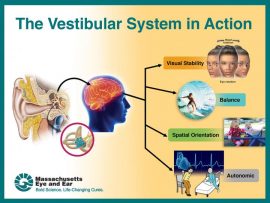
The vestibular system has three main functions: 1) Gaze stabilization which refers to ocular stability. This keeps images stable by moving eyes in response to head movements. 2) Postural control which detects position and movement of head in space; along with sensory and proprioceptive systems. 3) Perception of motion which helps to distinguish eye movements from head movements (internal) and head movements from exocentric (environmental) movements. The vestibular nuclei, cerebellum and reticular formation all receive input form visual and somatosensory systems. The output form these areas influence oculomotor control and spinal motor control. The central pathways for these systems are separate, therefore, both systems are examined and treated separately.

A vestibular exam will typically consist of:
• Acquiring a history
• CN testing
• Eye head coordination
• Positional testing
• Postural control
• Functional testing
• Locomotion

Other assessments include: Visual vertigo analog scale (VVAS)
Dizziness Handicap inventory/index (DHI)
Activity-specific Balance Confidence (ABC) Scale

Clinical decision-making model and differential diagnosis includes:
• Acute symptoms – Possible BPPV, labyrithinitis, stroke , fall and concussion
• Episodic – possible Meneire’s, postural hypotension
• Chronic – Possible Mal debarquement, hair cell loss, aging, long term CNS injury

Exam:
By taking our clients through various positional changes and movements of the head, we can elicit symptoms and this will help to determine where the lesion/dysfunction is originating and lead us to the best protocols to reduce symptoms. Following the steps below, one can observe ocular movements and fluidity of movements, lag in response to positional changes or presence of nystagmus (ticking of eye movements horizontally or vertically). Always take note of direction of the “beat”/tick as well as how long it lasts. Always assess the client’s perception of severity of vertigo on 0-5 scale with 5 being severe.

Visual field deficits: Normal: superior 60 deg; inferior 75 deg; tamporal 100 deg; nasal 60 deg.
Eye / head coordination: Eye range of motion – rectangular; eye coordinated, conjugate motion with head steady.
Smooth pursuit – smooth eye movement at less than 60 degrees per second; eyes tracking moving object
Saccadic eye movements – rapid eye movements between two targets
Vestibular Ocular Reflex (VOR) – gaze stability during rapid head movement
In-phase – eyes fixed on object; heading moving
Cervical Ocular reflex (COR) – rotation of body under head – keeping head stationary
Gaze Stabilization – Optokinetic system
• Combination of saccadic and smooth pursuit system
• Stimulated by repeated movements across a subject’s visual field (an object moving across the stationary visual field or by a person passing by a stationary visual field)
Nystagmus – non-voluntary rhythmic oscillation of eyes; fast and slow components beating in opposite directions; named by the fast component.
• Pathological nystagmus – appear with or without external stimulation in patients with vestibular disorders
• Spontaneous – present with head erect and gaze centered
• Positional – induced with changes in head position
• Gaze evoked – induced by change in eye position
Peripheral Vestibular lesion:
• Jerk nystagmus (named for the fast component: away from the lesion: either up or down beating) Side of lesion is opposite the quick motion (jerk)
• Pendular nystagmus (right or left beating; or up or down beating)
• Rotary nystagmus (named for direction of spin e.g. Upward and rotary)
• Result of asymmetry of right and left vestibular systems.

Head thrust test (HTT) or head impulse test (HIT) – clear neck AROM and carotid artery first
• Fixation on near target then far target ( 6 ft away)
o Slow head movements
o Fast head movements

• Watch for saccades – re-fixate on target (nose)
o Peripheral or central vestibular lesion – unable to maintain gaze
o Bilateral peripheral vestibular lesion – re-fixation to both sides

Head Shaking nystagmus test
• Eyes closed; head tilted downward to 30 deg (places horizontal canals // to ground)
• Turn head passively side to side 20x (2Hz)
• Check for nystagmus using frenzel lenses
o Unilateral peripheral lesion – asymmetrical nystagmus – slow phase toward involved (hypo-functioning) side
o Normal – no nystagmus
o Central lesion – vertical nystagmus

Clinical dynamic visual acuity test (DVAT-N)
• Measures functional VOR or ability for person to stabilize gaze during head movement
• Read visual acuity chart on wall 4 m away
o Lowest line read seated
o Lowest line read while head is passively oscillated in horizontal direction at 2Hz
*Vestibular hypofunction of >3 line change in visual acuity
Monofilament testing plantar sensation
Sensory levels 1= 1g Normal sensation
2= 10 g Protective sensation
3= 75g Loss of protective sensation
4= No perception
Motion Sensitivity quotient (MSQ) – Measures individual response to positional changes. (I.e. quickly supine to side lying R and L; supine to sit and return; wait for response
• Establish baseline of symptoms of vertigo/dizziness, nystagmus at rest
• Monitor symptoms of vertigo/nystagmus
Dix-Hallpike Maneuver
• Quick movement from sitting to sidelying with head rotated 30 deg away from downside ear
• Caution: check for neck AROM and vertebral artery prior to any quick motions of neck on older people
• Watch for nystagmus and direction
• Record duration and intensity of nystagmus and vertigo 0 (none) – 5 (severe)
o Nystagmus directional perponderance:
 Horizontal canal – nystagmus will occur with fast component toward the floor (Horizontal geotropic meaning, toward the earth) or Ageotropic – away from the earth
 Anterior canal – torsional and DB (down beat)
 Posterior canal – torsional and UB (upbeat)
The most common peripheral lesion is BPPV of the posterior canal. With testing, one will typically see and upbeating, torsional nystagmus. (There may be a 30 second delay in nystagmus). Short term is <2 minutes if BPPV. This will usually improve in one treatment. Allow rest and re-test for symptoms.

Evaluation and interpretation of findings
With evaluation, Peripheral lesions/dysfunctions will present with BPPV, nystagmus, short duration vertigo, possible hearing loss and/or tinnitus. While Central dysfunctional will present with head trauma, concussion, stroke, MS. Symptoms will include nystagmus vertically, long lasting, lateropulsion and head tilt.

Mechanical Dysfunctions to look for
• Benign Proxysmal Positional Vertigo (BPPV), with typically be a result of cupulolithiasis or canalithiasis. Symptoms will typically include vertigo with changes in head position, nausea with/without vomiting, disequilibrium
• Right Posterior Cupulolithiasis will typically present with a persistent upbeat nystagmus and right torsion. Canalithiasis will typically present with transient upbeat and right torsion.
• Left anterior Cupulolithiasis will typically present with persistent downbeat and right torsion. Canalithiasis will typically present with transient downbeat and right torsion.
• Horizontal cupulolithiasis will typically present with persistent upbeat nystagmus, while canalithiasis will typically present with downbeat nystagmus

Identification of Semicircular Canal if peripheral lesion:

Canal Involvement Primary nystagmus: right Hallpike-Dix Reversal nystagmus: Right Hallpike-Dix Nystagmus: return to sitting
Right posterior Upbeat and right-ward torsion downbeat and left-ward torsion Downbeat
Right Anterior Downbeat and right torsion Upbeat and left torsion upbeat
Horizontal Horizontal opposite horizontal direction opposite horizontal direction
Left Anterior Downbeat and left-ward tornsion Upbeat and right-ward torsion upbeat

Treatment
In most instances, the techniques presented below will significantly decrease or stop symptoms within 1-2 treatments. Once, vertigo symptoms are addressed, balance, advanced gait and strengthening can be addressed as well as accommodation techniques which will become more complicated/advanced as the client accommodates. These will involve increasing eye, head and body movement as the client improves.

Canalith Repositioning Technique: CRT-Posterior SSC (Canalithesis BPPV) Treating Left side.
• Turn head left 30 deg from midline
• Maintaining 30 deg head turn, move to supine position
• While in supine, turn head to opposite side (right), 30 deg from midline
• Have client roll to onto right side while still maintaining 30 deg head turn to the right
• Transition back to upright position while maintaining 30 deg head rotation

Cupulolithesis (for posterior SSC) Treating Right side
• With client in sitting position, rotate head 30 deg right
• Move to side lying right, maintaining head rotation
• Keeping head at 30 deg rotation from midline, have client sit up and move to side lying Left
• Then move back to sitting. Head will be in original 30 deg to Right.
*once head rotation 30 deg from midline is achieved initially in sitting, this head position is maintained throughout the maneuver.

Cupulolighesis BPPV (for anterior SSC) Treating right side
• Have client in sitting position, turn head 30 deg to left
• Transition to side lying right (with head maintained in 30 deg rotation – head rotation will be up toward ceiling)
• Then move to left side lying while maintaining rotation 30 deg to left – head rotation will be toward the floor)
• Then have client move back to upright sitting, maintaining head rotation to left 30 deg.

Horizontal Canals – Roll Test
• With pt in supine, to test right side, have client rotate head to the right
• To test left side, have client rotate head to the left
*Alternative position can be performed with client’s head on a pillow or wedge

Canalith repositioning technique: CRT – Horizontal SSC
• Start with client in side lying with “bad” ear down
• Roll to supine
• Then move to opposite side lying position with “bad” ear up
• Then move to quadruped position with head // to the floor

Habituation Techniques:
• General habituation technique for posterior SSC BPPV. In sitting position, rotate head 30 deg Left, move to side lying left, then back to sitting, repeat on opposite side. Remain in position 30 sec or until vertigo stops. Perform 10-20 x TID.
• Gaze stability – looking at a fixed object and turning the head slowly from side to side.
o Turning body under the head with head fixed
o Body/head fixed looking at a central object, moving eyes only look to objects above, below, laterally and diagonal to central object/point
o Balance activities – stepping over objects, around objects.
o Gait activities with head movement

The activities outlined above are just a starting point. Be creative and always create an individualized program for each client based on symptoms, persistence of symptoms and each client’s specific deficits, their specific goals and activities and hobbies each client wants to return to.
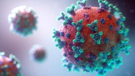
Think Thin! Collaboration in Flagstone

Morgan Nebo
Aggie Smith

Morgan Nebo, Dietary Supervisor at Victoria Post-Acute, and Aggie Smith, Flagstone Dietitian, provided a great presentation to the Flagstone DORs and SLPs on best practices for therapy and dietary collaboration with emphasis on ThinkThin!

Key takeaways from this presentation:
1. CMS — Quality of Care Intent: “To the extent possible, MAINTAIN or IMPROVE before complications arise.”

● Resident HYDRATION is the key (and the challenge) with thickened liquids.
● F692, Quality of Care Nutrition and Hydration, requires that a resident is offered sufficient fluid intake to maintain proper hydration and health.
● As a general rule, most residents will require 1500–2000 cc daily under “normal” circumstances — SLP collaboration.

2. Nutritional Assessment and Risk Identification: Collaboration with SLP and Food/Nutrition to assess a variety of areas.
3. Become familiar with using the information in PCC: i.e., Nutritional Assessment UDA along with CNA input/output reporting.
4. Collaboration is super important during quarterly assessments
5. Become familiar with the facility menu system in the affiliate you are servicing. Review the diet manual standards and follow facility nomenclature. If a facility-wide change is needed, collaborate!

The training was so successful, the LMS team is working on a recording so everyone in the organization can benefit from the great information and have continued collaboration for Think Thin!

Sharing Vital Sign Tips

By Dawn Thompson, DOR, Victoria Post Acute Care, El Cajon, CA

Over the last several months, Victoria Post Acute Care [VPAC] has honed in on vital sign monitoring as part of our daily practice in both nursing and therapy. I’ve been nominated to lead our vital signs committee, and during the next few weeks I’d like to share some of the best practices and education that assisted us through COVID and landed VPAC Vital Sign Grand Champions.

We’ll start with the vital sign we’re all monitoring daily, body temperature. After 8 months of daily screening, you’ve probably noticed a pattern and can guess your morning temperature within a few tenths of a degree; you’ve established your baseline temperature.

Do you know your residents’ baseline temperature? Would you be able to identify a fever and assist in early recognition of an infection?

Regularly measured body temperature is used to establish a baseline temperature and define the threshold temperature for fever. The elderly are noted with a lower baseline body temperature and a shift of 2.4*F above baseline is a fever, even when falling below definition of a fever at 100.4*F.

At VPAC, once a baseline temperature has been established, it is then added in special instructions in the Care Profile in PCC [See photo below]. This baseline temperature can then be used to assist in identifying any potential infection or COC with our residents.

Please don’t hesitate to reach out with any questions. (DawnThompson@ensignservices.net).


Making Vital Signs Vital

By Tamala Sammons, M.A., CCC-SLP, Therapy Resource
We have been focused on why it is so important to measure vital signs as part of our clinical practice. Not only does the data help us with early detection of sepsis, identifying patient instability, having comparative baselines during exercise, and knowing when to stop an activity, but it also helps us make better clinical decisions around patient care. Now, therapy teams need to also focus on capturing measured vital signs into our daily documentation practices.

Measuring and documenting vital signs starts at the evaluation to ensure treatment plans are designed to address challenges with varying diagnoses and to ensure we provide interventions accordingly.

Next, vital signs are measured during treatment sessions to support decisions around interventions being provided. The key for us as therapists is to capture the data from vital signs as a guide to what interventions we will provide, or stop providing. In other words, we need to do more than simply take and record vitals. We need to use vital sign data as tools to make clinical decisions.

For example:

● Mr. Smith has O2 weaning as a goal. OT is documenting patient is SOB during activity; however, no vitals were recorded at evaluation or in TENs. Data was only entered into PCC. Here is why capturing vitals in our clinical documentation is also needed: Documentation is further enhanced when the OT documents how many liters the patient is on and levels of O2 before the activity, five minutes into the activity, and after the activity. This documentation is specific to the therapy session and needs to be recorded in the TENs as it supports what physical activity the patient can tolerate as part of decisions around O2 weaning and overall improvement with ADLs.

● Mr. Jones has precautions with BP risk identified on the PT evaluation. However, upon review of daily TENs, BP is not captured in the documentation pre-, five minutes into, and post-exercise. BP was entered into PCC, which is great for simply recording data. However, using the data in a meaningful way in our TENs supports the clinical judgment of a therapist (i.e., what decisions during treatment were made based on BP readings?) and further supports medical necessity for care.

● Mr. Romero had a CHF exacerbation and the hospital record noted he has 45% ejection fraction and he has SOB with walking greater than 75 feet. Documented vitals are paramount to ensure that his treatment with functional activities or prescribed exercise are keeping his HR between 25-35% of his target HR.

Other Examples:

● Pt. “V” has SOB and needs to rest frequently. What do the vital signs tell us and do we change direction in treatment due to those measures?

● Pt. “I” is on 2L O2. PT is working on ther ex and gait. Does O2 change with exercise? How long does it take to get them to recover?

● Pt. “T” is medically complex and post-septic. What do we know about pts who have been septic? How are we ensuring we are monitoring for s/s of sepsis while in therapy? What vitals are recorded in our documentation to support our clinical treatment decisions?

● Pt. “A” is doing breathing exercises with SLP due to COPD. What is the patient’s respiratory rate pre-, during, and post-breathing exercises? What are the O2 levels? How do we know the interventions are working?

● Pt. “L” is a cardiac patient. How are vitals documented during treatment sessions to support exercise prescription? How do we know our plan is working?

Please continue to work with your teams to not only measure vitals, but more importantly, integrate the data into evaluation and treatment documentation to support skilled intervention decisions, capture clinical judgement, and demonstrate medical necessity of our services.


Therapy to ED Leadership

By Brian del Poso, OTR/L, CHC, RAC-CT, Therapy Resource
Next up in the series of interviews of our former DORs turned ED is the one and only Amy Gutierrez! She is not only a former DOR, but served as a Therapy Resource as well. Amy is currently the ED at Treasure Hills in Keystone. She was kind enough to share some of her thoughts with us:

As a DOR/Resource, you were in a good place in your career. What kinds of things were you thinking about when the thought of being an ED came up?
Jon (Anderson) was actually the one that brought it up in October of 2018 at the Leadership Summit meeting. Prior to that, I never really wanted the responsibility lol! I suppose that was when the seed was planted. Throughout that time period I considered taking my boards to becoming an Admin, I started asking questions to the other administrators. One of the most frequent questions I had was, “Is it fulfilling?” As a therapist and a resource, I know we make a difference and felt I had a purpose. I didn’t know if I would have the same sense of fulfilment as an ED. I was naïve to think that it wouldn’t be.

How did you come to the decision to push forward into the AIT/CIT program?
To be honest, I kind of jumped in. The position was offered to me at the beginning of October with the acquisition scheduled to happen in November. At that time, I was still in my Hybrid role as a DOR and Resource. I was fortunate enough to attend an AIT boot camp, which helped to solidify the decision I made in becoming an ED. As an Administrator, we are given an opportunity to change the lives of many. And where we do that as therapists and Resources, we have the opportunity to do it on a much broader stage. All of those little changes we want to make, or we wish the Admin would do, is now on us. I learned rather quickly nothing is ever as easy as it appears. We are entrusted with so much, at times it could feel overwhelming, but it’s in those moments you begin to see growth as a leader.

What do you think is the most important characteristic of a successful leader?
This is a hard question to answer. I admire so many of our own leaders but for different reasons. One of the most common traits they all have is their openness to give and receive feedback. They surround themselves with the right people. They share what they know and want every single person around them to be just as successful.

If you were to talk to a therapist about the ED role in general, what is the most important thing you would talk to them about?
Being open and honest about the things you don’t know. Ask questions, lots of them. I find myself calling my partners and resources multiple times a day. It’s OK not to have the answers. One of my mentors shared that with me early on. And it has saved me a million times over.

Any other thoughts for a therapist who might be thinking about becoming an ED?
You have to be ready for anything. I have been in my role for eight months now, and I can honestly say, I never expected to be an Administrator under the world’s current circumstances. But you do everything in your power for the people under your leadership and those entrusted to your care. Times and positions like these are what therapists are made for.

Think Thin! The Path to Thin Liquids

By Tamala Sammons, M.A., CCC-SLP, Therapy Resource

A new clinical campaign for our SLPs and IDT is the “Think Thin! A Path to Thin Liquids” approach. There is so much supporting evidence that promotes thin liquids over thickened liquids. When thick liquids are needed, then we need to consider utilizing the free water protocol.


Reasons to Think Thin:

Preventing Dehydration: Dehydration can lead to a variety of negative health consequences including:
• Changes in drug effects
• Infections
• Poor wound healing
• UTI’s
• Confusion
• Constipation
• Altered cardiac function
• Declining nutritional intake

Improving Quality of Life:
Traditional thought holds that aspiration of any material into the lungs can lead to aspiration pneumonia so many patients who have difficulty swallowing are placed on diet restrictions that avoid thin liquids.
However, a confounding evidence in the literature suggests that pulmonary aspiration of differing materials may not present an equal risk for the development of aspiration pneumonia. Aspiration will result in pneumonia only if the aspirated material is pathogenic to the lungs and the host resistance to the aspirated material is compromised. Research also discovered: “The risk of developing aspiration pneumonia was significantly greater if thick liquid or more solid consistencies were aspirated.” (Holas, DePippo, & Redding, 1994)

Being able to have Thin Water: Free Water Protocol
If a patient must be on a thickened liquid for any duration of time, research using a free water protocol found that fewer residents had UTI’s and dehydration and that when paired with proper positioning and oral care, there were no incidents of aspiration. Additionally, providing patients with thin water:
• Improves quality of life
• Improves Resident satisfaction with meals and less reports of thirst (Over 35% of patients are noncompliant with thickened liquids)
• Decreases risk of dehydration, UTI’s and pneumonia

Additional training information and materials will be coming over the next few weeks as we work to Think Thin!